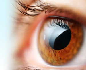
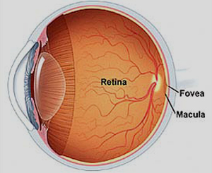


What is a retinal detachment?
A retinal detachment refers to a situation in which retina is separated from its attachments to underlying tissue within eye. Mainly retinal detachments are an outcome of a retinal tear or break. These retinal breaks are caused due to vitreous gel pulls that occurs mainly in peripheral parts of retina. The vitreous is a clear gel of which two-third is filled inside eye and it resides in front of retina. When vitreous gel gets liquidified, the retina becomes weak and as a result it will tear or break down. Most retinal breaks don’t occur as a result of injury. Sometimes retinal tears are accompanied by bleeding. Vitreous gets separated from retina in cases of many people when they get older. However, only a minute percentage of these vitreous separations result in retinal tears.
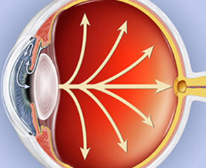
Once retina has become weak and gets torn, liquid from the vitreous gel collects behind retina. The upsurge of fluid behind retina detaches retina from the back of eye. As more and more of liquid vitreous gel collects behind retina, the risk of retinal detachment can progress and even involve the entire retina, to a situation of total retinal detachment. In many cases, it has been seen that a retinal detachment only affects one eye at a time.
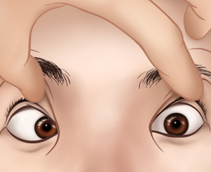
What are the symptoms and signs of retinal detachment?
The initial symptoms of a retinal detachment are flashing lights and floaters. If anyone is experiencing these symptoms, then he should see an eye doctor for a retinal exam. Sometimes symptoms of flashing lights and floaters are unassociated with a tear or detachment and can simply result because of separation of vitreous gel from retina. This condition is often known as posterior vitreous detachment (PVD). The floaters are caused because of condensations in vitreous gel which are frequently described by patients as strands, spots, or little flies. If the patient is experiencing a shadow or a shade which affects any part of his vision, this type of situation indicates that a retinal tear has progressed into a detached retina. In such circumstances, one should right away consult or seek an advice from eye doctor.
What are the treatments of retinal detachment?
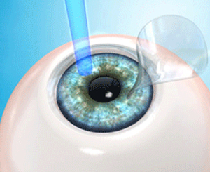
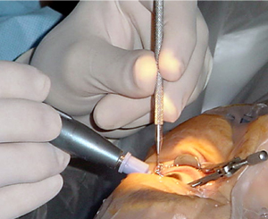
Usually surgerical therapies are used for curing retinal detachment disease. Some of these therapies are:
Scleral Buckling: This is the most used therapy for curing retinal disease. This surgery requires hospitalization of patient in which the patient is given local anesthesia before proceeding with this surgery. Cryotherapy is used to block retinal tears which holds retinal tear against the sclera. In certain cases surgeon paces air bubble inside the vitreous cavity in order to drain fluid underneath the retina. The success rate of this type of surgery is about 90-95% approximately.
Pneumo-retinopaxy: In this surgery you will be instructed to keep your head in a specific position by which gas bubble blocks and seal s retinal tear. As a result of which fluid circulation stops which results in improved sight vision.