


Examine the corneal diseases with the help of Specular Microscopy
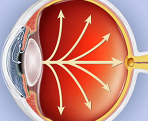
Specular Microscopy is non-invasive method that is used for examining, monitoring and documenting the quality and counts of the endothelial cells which lines up the corneal back. This method is quite useful in determining the various corneal diseases that can affect the normal eye-vision. This is done because at the age of 10 years, an individual carries healthy cornea which is comprised of almost 3500 cells that too within each mm square. But with normal process of ageing i.e. by the age of 60, these cells keep on degrading in number and it reaches to 2500 cells per mm square.
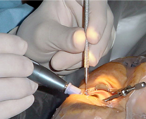
This procedure is done by the ophthalmic laser technologist for screening the distinct corneal diseases that includes Keratoconus, traumatic injury, Fuch’s Dystrophy, etc. It is also very effective method for examining and evaluating the localized haze, edema or trauma inside the cornea that occurs due to the IOL lenses and contact lenses. This test takes only 15 to 20 minutes and it do not require any special preparation for it. Specular Microscopy provides simple and rapid way for obtaining accurate information about the corneal back which is not possible with the topography or standard biomicroscopy.
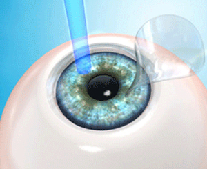
This procedure involves the usage of a special instrument called as specular microscope that permits the photographer as well as the doctor to visualize and record cells of cornea. It does not require any cut or incision; it directly touches the front of the eye or else the cornea. It requires the help of a bright image which is formed when the position of light is set at the right angle and since the corneal tissues are clear, hence only a little amount of the light returns back for the photography.